

热门关键词:
{pboot:nav num=12 parent=5}
Low-Temp Plasma Radiofrequency Ablation System|
Dynamic System (Grinding Drills Suits)
NEWS
Training & Education
contact
立即咨询
热门关键词:
{pboot:nav num=12 parent=5}
Dynamic System (Grinding Drills Suits)|
Active Biological Bone
NEWS
Training & Education
contact
立即咨询
热门关键词:
{pboot:nav num=12 parent=5}
Active Biological Bone|
Unilateral Biportal Endoscopic (UBE) Surgical Instrument
NEWS
Training & Education
contact
立即咨询
热门关键词:
热门关键词:
热门关键词:
{pboot:nav num=12 parent=5}
Sports Medicine Surgical Instruments|
关闭
![[x]](/template/default/img/closeicon1.png)

News center
Spinal minimally invasive foraminoscopy technology, small incision to solve big problems!
Author:COMFIER MEDICAL
Spinal minimally invasive foraminoscopy technology, small incision to solve big problems!
A foraminoscope is similar to a spinal endoscope in that a lighted tube is inserted into the foramina from either the side or the back of the patient's body (either flat or oblique) to perform surgery in the safe working triangle.
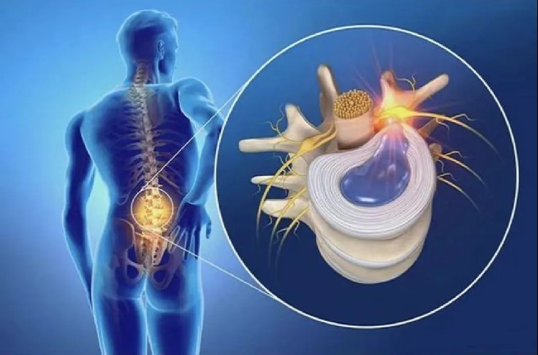

The protrusion of nucleus pulposus, nerve root, dural sac and proliferating bone tissue can be clearly seen under the direct view of the endoscope. Then various types of grasping pliers are used to remove the protrusion tissue, bone removal under the microscope, and radiofRF electrodes are used to repair the damaged annulus.
The surgical trauma is very small, like the size of a soybean kernel, the bleeding is less than 20ml, and the postoperative suture is only 1 needle, which is the minimally invasive treatment of the intervertebral disc herniation in the similar surgery.
Advantages of foraminoscopy
The incision is only 6-8mm, which has little impact on the integrity and stability of the spine and is easily accepted by patients

Traditional open surgery incision is large, trauma is large, affect the appearance

The incision of foraminoscope is small and the appearance is more beautiful
Visual operation, clear vision, accurate positioning, avoid damage to nerves and blood vessels, local anesthesia operation, the entire operation of patients fully awake, low risk of surgery.

Back to list

分享:
Previous:Several thoughts on the service life of equipment medical devices
Next:None
News recommend
Spinal minimally invasive foraminoscopy technology, small incision to so···
Application of Endoscopy in Spinal Surgery
Several thoughts on the service life of equipment medical devices
Several thoughts on the service life of equipment medical devices
Several thoughts on the service life of equipment medical devices